Petelle2

Tracking chicks to understand individual movement phenotypes and their corresponding welfare outcomes

Tracking chicks to understand individual movement phenotypes and their corresponding welfare outcomes

Our research group focuses on the health and welfare of poultry and rabbits as it relates to their housing. To this end, one of the main themes in our research is individual variation in movement and space use within a commercial setting. Our past research shows that individuals are consistent in how they move throughout the aviary, and that individuals are distinctly different from one another. For example, some individuals are very active, moving throughout the different levels of the aviary rapidly throughout the day, while others tend to stay on one level for most of the day. However, we don’t know when distinct individual variation emerges or how it changes across the lifetime of the individual.

With the generous support of the UniBern Forschungsstiftung, we were able to purchase additional radio frequency identification (RFID) antennas to install in our rearing barn to gather positional data of chicks at one day of age. From this data, we will assess whether individuals differ in their space use right after hatch or whether differences develop slowly over the rearing period. We can then link these movement differences with overall health and welfare across their lives.

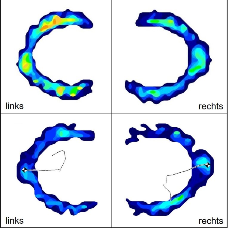
At present, we have already carried out a pilot study in our experimental barn (Figure 1) that demonstrates we are able to obtain data from chicks and that movement between chicks across the first weeks seem to be consistently different, however our validation is ongoing. We also recently installed antennas in two pens in our rearing barn and started tracking 600 chicks (300/pen) to determine movement differences in a commercial setting (Figure 2).

Drs. Michael Toscano and Matthew Petelle

ZTHZ – Center for Proper Housing: Poultry and Rabbits

VPHI – Veterinary Public Health Insitute


Figure 1: Fifteen chicks in one of our pilot pens. Each chick is outfitted with an RFID wing tag to monitor their location within the pen.
Figure 2: One day old chick with RFID tag in commercial rearing pen. Antennas are under the brown chick paper.
Jeckelmann1 - Kopie

Glow discharge apparatus for high quality and reproducible cryo-electron microscopy specimen preparation

Glow discharge apparatus for high quality and reproducible cryo-electron microscopy specimen preparation

Cryogenic electron microscopy (cryo-EM) has become the preferred method for determining the atomic-resolution structures of macromolecules 1. The preparation of cryo-EM specimens is a multi-step procedure, typically divided into four main stages: (i) glow-discharging the cryo-EM grids, (ii) applying the macromolecular sample to the grid, (iii) blotting off excess protein solution, and (iv) vitrifying the specimen in a cryogenic liquid. Specialized equipment is employed in steps (iii) and (iv) to meticulously control these processes. The importance of vitrification in cryo-EM grid preparation was highlighted when the Swiss researcher Jacques Dubochet received the Nobel Prize in Chemistry in 2017 for his pioneering work in this area (https://www.nobelprize.org/prizes/chemistry/2017/summary/).

Cryo-EM grids used for high-resolution protein structure determination are typically made of copper or gold with a holey carbon film attached on one side. Chemically, this surface is hydrophobic and would naturally repel hydrophilic protein samples. Therefore, cryo-EM grids must be rendered hydrophilic through a process called glow-discharge. Since the glow-discharging procedure significantly impacts protein absorption behavior on cryo-EM grids, precise control of this process is essential. With the funding acquired through the UniBern Forschungsstiftungs grant, we obtained a Dual-Chamber PELCO easiGlow™ glow-discharging device, which allows for controlled glow-discharging and ensures a more reliable and reproducible production of cryo-EM specimens (Figure 1A).

The PELCO easiGlow™ glow-discharging device was installed in the sample preparation room of the structural biological branch of the Microscopy Imaging Center of the University of Bern (MIC, https://www.mic.unibe.ch/). Since its installation, this device has been used to produce cryo-EM specimens that are analyzed on the MIC’s high-end electron microscope Titan KRIOS G4. The use of the PELCO easiGlow™ device has already proven invaluable to our research, enabling our group to recently publish the high-resolution structures of (i) the bacterial green-light-absorbing proton pump proteorhodopsin (GPR) 2 (Figure 1B) and (ii) the bacterial glucose transporter IIC(B) 3 (Figure 1C). Moreover, the PELCO easiGlow™ glow-discharging device holds significant value for all MIC users at the University of Bern.

Jean-Marc Jeckelmann, PhD
Institute of Biochemistry and Molecular Medicine

Links:

1             Guaita, M., Watters, S. C. & Loerch, S. Recent advances and current trends in cryo-electron microscopy. Curr. Opin. Struct. Biol. 77, 102484 (2022). https://doi.org/10.1016/j.sbi.2022.102484

2             Hirschi, S. et al. Structural insights into the mechanism and dynamics of proteorhodopsin biogenesis and retinal scavenging. Nature Commun. 15, 6950 (2024). https://doi.org/10.1038/s41467-024-50960-3

3             Roth, P. et al. Structure and mechanism of a phosphotransferase system glucose transporter. Nature Commun. 15, 7992 (2024). https://doi.org/10.1038/s41467-024-52100-3


Raissig2

A hand-held porometer for high-throughput phenotyping of plant-atmosphere gas exchange

A hand-held porometer for high-throughput phenotyping of plant-atmosphere gas exchange in grasses

Land plants must balance water vapour loss through leaves with efficient carbon dioxide (CO2) uptake for photosynthesis. Specialised “breathing pores” on leaves called stomata can open and close to minimise water loss and maximise CO2 uptake. Thus, plants with efficient and fast stomatal pores are likely more resilient to the upcoming, climate-change-induced drought and heat periods.

The ”Stomatal Biology” group at the Institute for Plant Sciences is interested in how different stomatal morphologies affect gas exchange and how we can bioengineer stomatal form to prepare plants for the upcoming climatic challenges. We primarily work with grasses, which form morphologically innovative stomata with very rapid opening and closing dynamics. The rapid stomata of grasses contribute to the high water-use efficiency of grasses and their evolutionary success. Nowadays, grasses dominate many natural and agricultural ecosystems and our most important food crops like maize, rice and wheat are all grasses.

Gas exchange measurements are either very time-consuming and laborious or rather inaccurate. With the help of the UniBern Forschungsstiftung, we were able to acquire the hand-help porometer LI-600N, which allows for rapid and highly accurate measurements of steady-state gas exchange, while simultaneously assessing photosynthetic capacity. Therefore, the LI-600N will enable us to perform high-throughput screens of large populations of different grasses or grass genotypes. This will identify species that show a high photosynthetic efficiency, which is relevant for yield, yet low stomatal conductance, which is relevant for water-stress resilience.

Prof. Dr. Michael T. Raissig
Institute of Plant Sciences (IPS)

https://raissiglab.org/
https://www.ips.unibe.ch/

Raissig1
Figure 1: The measuring head of the porometer LI-600N for narrow leaves (left) and LI-600 for broad leaves (right).
Figure 2. The LI-600N in action measuring steady-state stomatal conductance and photosynthetic efficiency (with the red-light pulse) of the wild grass Brachypodium distachyon.
Leidel1

Using big data for the analysis of cellular translational control

Using big data for the analysis of cellular translational control

Protein synthesis is essential for any living organism. However, how the dynamics of mRNA translation affect the formation of functional proteins is poorly understood. To gain new insights into the relationship between translation dynamics and protein folding, we analyze mutants that show codon-specific translation defects.

We analyze these mutants in detail using a variety of omics techniques such as ribosome profiling or pulse-chase proteomics. As all these methods generate large amounts of data, it is essential to be able to store and handle such large datasets. With the funding we received from the UniBern Forschungsstiftung, we purchased an extension to our redundant NAS storage system and a new analysis server. We have already used it to characterize key enzymes in translation dynamics (Wu et al., bioRxiv 2024; Lin et al., Mol Cell 2024).


The next goal is to develop machine learning strategies to analyze and integrate these datasets. However, the extension of our NAS system, combined with the purchase of a new analysis server, has already led to exciting new discoveries and will continue to do so in the future.

Prof. Dr. Sebastian A. LEIDEL

Department of Chemistry, Biochemistry and Pharmaceutical Sciences

Links:

– Wu et al., BioRxiv 2024: DOI: 10.1101/2024.02.27.582385

www.biorxiv.org/content/10.1101/2024.02.27.582385v2

– Lin et al., Molecular Cell 2024: DOI: 10.1016/j.molcel.2024.06.013

https://doi.org/10.1016/j.molcel.2024.06.013

Karousis22

Development of an in vitro translation-based screening system to identify mRNA translation inhibitors

Development of an in vitro translation-based screening system to identify mRNA translation inhibitors

The goal of our project is to develop a platform that allows the identification of small molecules that inhibit translation in human cells using cell-free lysates. The lysates are also used for studying the mode of action of proteins from coronaviruses (2,3).

With the funding that was acquired thanks to the UniBern Forschungsstiftungs grant we obtained a shaking incubator device that allows us to produce ample amounts of cells and lysates for our screening purposes, based on a previously published protocol (1).


Evangelos D. Karousis, PhD
Dept. of Chemistry, Biochemistry and Pharmaceutical Sciences

Fördersumme 2024

Die Fördersumme unserer Stiftung zugunsten der Forschenden der Universität Bern beträgt im Jahr 2024 insgesamt CHF 359’561.–. Weiter unten auf dieser Seite ist die Liste der bewilligten Gesuche – geordnet nach Fakultäten – einsehbar. Die Antragssumme belief sich auf total CHF 467’284.–, verteilt auf 46 Gesuche.


Die Förderbeiträge werden finanziert aus dem allgemeinen Betriebsfonds unserer Stiftung, der Zuwendung der IMG Stiftung, dem Beitrag aus dem BEKB Förderfonds, der Zuwendung für Nachwuchsforschende und dem Legat Schwemer. Die Stiftung hat damit aktuell die Möglichkeit, über insgesamt fünf Fördergefässe Beiträge an die Forschenden auszuschütten. Mehr Informationen dazu finden Sie hier. Der allgemeine Betriebsfonds finanziert sich durch den Ertrag der Wertschriften und Spenden von Privatpersonen, Stiftungen und Firmen, mehrheitlich aus dem Kanton Bern.

Bild_2023-11-07_223750511

Cardiac channelosomes: focus on the sodium channel Nav1.5

Cardiac channelosomes: focus on the sodium channel Nav1.5

Numerous human diseases are caused by genetic or acquired ion channel dysfunction called channelopathies. Ion channels are membrane proteins allowing the passage of ions. The cardiac sodium ion channel Nav1.5, which gene is SCN5A, plays a pivotal role in such disorders. Nav1.5 channels are mainly expressed in the heart. Several hundred genetic variants in SCN5A were found in patients with a broad spectrum of cardiac manifestations, such as LQTS type 3, BrS, atrial fibrillation, and dilated cardiomyopathy. As with other proteins, the Nav1.5 channel interacts with many proteins, leading to its fine-tuning of expression, localization, and function. The work of our group is focused on deciphering which partner proteins interact with Nav1.5, leading to its proper function. The main technics used to perform such investigations are biochemical assays named co-immunoprecipitation (co-IP) experiments. However, the crucial step for this biochemical assay is the extraction of large proteins such as Nav1.5 α-subunit monomer (~220 KDa). In addition to their size, the embedment of those ion channels in a lipid bilayer, such as a plasma membrane, renders the extraction more difficult than cyto-soluble protein.

The homogenizer ‘Bioprep-24R’’ (ALLSHENG) that we were able to acquire, thanks to the UniBern Forschungsstiftungs’ grant, becomes essential for such investigation. The team members largely benefit from this new equipment to extract efficiently voltage-gated ion channels expressed in different organs and tissues. The homogenizer permits fast, effective, and reproducible homogenization, relating to the three-dimensional high-speed vibration and beating of grinding beads (glass beads, ceramic beads, steel balls, etc.).

We can now efficiently extract Nav1.5 channels from animal organs, a critical step to performing co-immunoprecipitation experiments.

PD Dr. Jean-Sébastien ROUGIER
Institute of Biochemistry and Molecular Medicine

Western blot showing the efficiency of Nav1.5 extraction from murine hearts using either the classical homogenization procedure or the Bioprep-24R.

The homogenizer ‘Bioprep-24R’’ (ALLSHENG) and western blot showing the expression of Nav1.5 channel exclusively in the murine heart (ventricles).


Karousis3

Production of translation-competent human cell lysates using dual centrifugation

Production of translation-competent human cell lysates using dual centrifugation

Protein synthesis is a central process in gene expression and the development of efficient in vitro translation systems has been the focus of scientific efforts for decades. The production of translation-competent lysates originating from human cells or tissues remains challenging, mainly due to the variability of cell lysis conditions.

With the funding that was acquired thanks to the UniBern Forschungsstiftungs grant we obtained a dual centrifugation device that allows for detergent-free cell lysis under controlled mechanical forces (published in Gurzeler et al., RNA biol., 2022).

We optimized the lysate preparation to yield cytoplasm-enriched extracts from human cells that efficiently translate mRNAs in a cap-dependent as well as in an IRES-mediated way. Using the derived lysates, we contributed to elucidating the role of Nsp1, a potent virulent factor produced during the early steps of infection by SARS-CoV-2 in human cells (Schubert, Karousis et al., Nature Struct. Mol. Biol, 2020) in collaboration with the group of Nenad Ban.

Additionally, we routinely use the technique now for immunoprecipitation experiments or for structural studies and we explore the potential for using it as a method of cell fractionation. The next goal is to develop a screening platform for human translation inhibitors based on in vitro translation. Therefore, the acquisition of this equipment led to the development of new exciting projects in the field of human translation.

Evangelos D. Karousis, PhD
Dept. of Chemistry, Biochemistry and Pharmaceutical Sciences

Die Projektförderung wurde ermöglicht durch einen Beitrag des BEKB Förderfonds