Refining quantitative sensory testing methods to assess chronic joint pain in horses
Refining quantitative sensory testing methods to assess chronic joint pain in horses
Our research group aims at improving pain recognition and treatment in animals. As animals cannot communicate verbally their feelings, it is of particular importance to rely on objective tools that allow measuring pain and its modulation in an objective way.
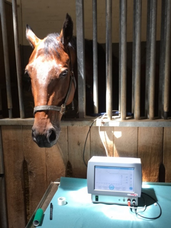
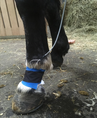
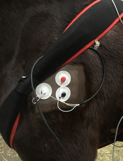
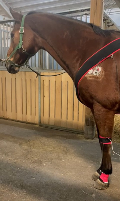
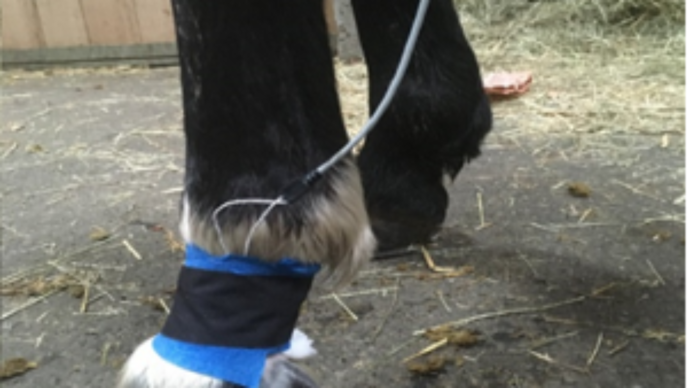
With the financial support of the UniBern Forschungsstiftung we were able to acquire a Dolosys Paintracker (Fig. 1), that allows a non-invasive, continuous determination of the «nociceptive withdrawal reflex» threshold (the pain threshold) in animals of different species. Through surface electrodes, electrical stimuli are applied to a peripheral nerve (Fig. 2) and the evoked muscular response is quantified by electromyography (Fig. 3 and 4). Based on predefined criteria, the software automatically detects the nociceptive threshold and tracks it over time. The automated continuous threshold tracking, compared to the classical methodology applied in past trials, opens new possibilities in several domain of pharmacological and clinical pain research.
At present, with the use of the Dolosys, we aim at developing a Conditioned Pain Modulation (CPM) paradigm that allows quantifying the extent of endogenous pain modulation in healthy horses. Later, the developed CPM tool will be applied to horses affected by chronic pain to establish whether impaired endogenous pain control substantially contributes to the observed clinical pain phenotype, as observed for several chronic pain conditions in humans. This might strongly influence our approach to therapy, increasing the chances of successful clinical outcomes and thus overall contributing to improved animal welfare.
Prof. Dr. Claudia Spadavecchia
Anaesthesiology and Pain Therapy Section Vetsuisse Faculty