"Specific effect of anaesthetic drugs on the cortical electroencephalographic activity."
Our group is performing research on anaesthetic and antinociceptive processes in animals. We have a solid expertise for applied veterinary neurophysiology.
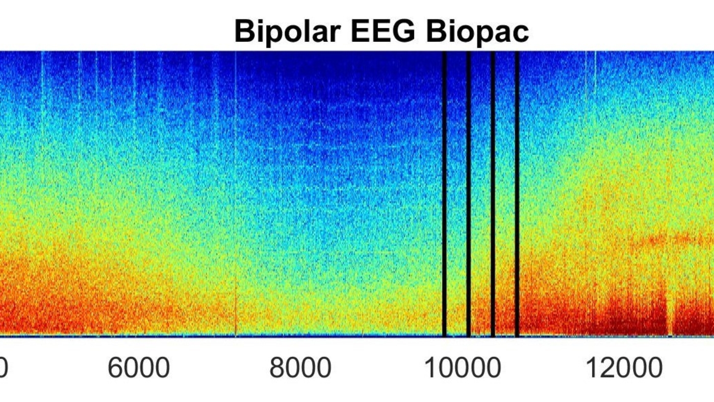
One of our current research focus is the investigation of the electroencephalographic (EEG) modulation from anaesthetics. Our aim is to characterize means of monitoring depression of consciousness (depth of anaesthesia) during general anaesthesia. Many studies have been performed in humans, and refinement of the methods is still ongoing. Recent developments include the evaluation of the color density spectral array from the frequency analysis of the EEG signal. Data in animals is very sparse.
A further output of our research is oriented towards a better understanding of mechanisms of unconsciousness during anaesthesia and compared physiology among animal species.
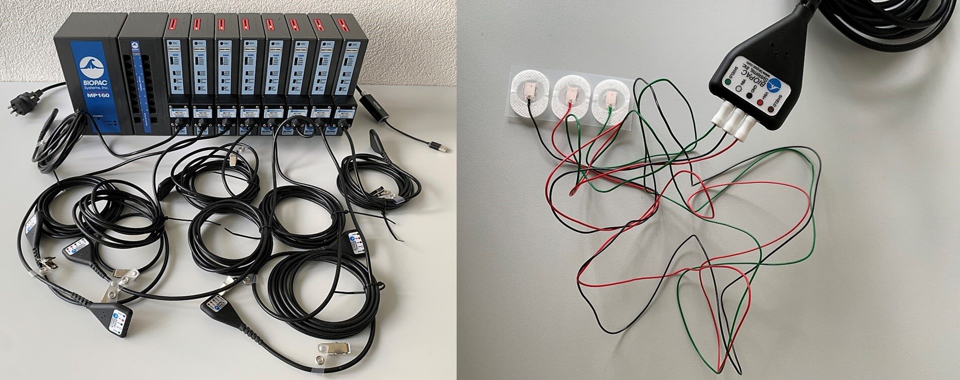
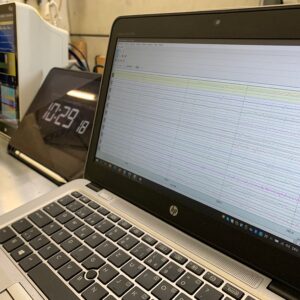
The present project is focusing on the modulation of the EEG activity by Propofol in pigs. Up-to-date equipment to capture and record the EEG in real-time was purchased including an experimental amplifier-recording unit (BioPac) and its software package (Acknowledge). The Berne University Research Foundation has awarded us a grant for 2/3 of the costs of the acquisition of the electroencephalographic (EEG) machine and the related software.
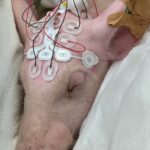
Electroencephalographic and electromyographic activity was collected in pigs receiving increasing dose of Propofol. Data were obtained from electrodes placed at the surface of the skin over the skull of the animal (frontal, temporal and caudal brain regions). Raw EEG data were processed to obtain power spectrum and a continuous time course of the density spectral array was evaluated, as well as other EEG-derived parameters (e.g. spectral edge frequencies, suppression ratio, frequency band ratios).
Specific patterns on the EEG signal could be investigated in relation to propofol administration and species-specific standardized anaesthetic signature observed and investigated.
Dr. med. Vet. Olivier Levionnois
Departement für Klinische Veterinärmedizin (DKV)
Klinische Anästhesie
Die Projektförderung wurde ermöglicht durch einen Beitrag des BEKB Förderfonds


